
Graft Dissection
Dr. Keene was the first doctor to introduce video magnification for hair restoration surgery. She first announced it at the International Society of Hair Restoration Surgery annual scientific meeting in Florida, 2002, and demonstrated it at the meeting in Rome, 2003. State of the art clinics have adapted this improved method of dissection.
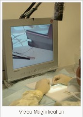
Research has shown that graft dissection using microscopic magnification reduces the incidence of transection, increasing graft yield during graft dissection. Table top microscopes have limited working distance and visual field, with limited range of motion for the users head and neck.
Dr. Keene sought a way to improve ergonomics and magnification over the commonly used microscopes used by hair transplant clinics. While seeking an ergonomically improved method of graft magnification, Dr. Keene worked with an engineer to co-develop the video magnification system, which not only offers ergonomic advantages, but also allows the doctor the opportunity to see every graft as it is being dissected. This also provides her the opportunity for maximum oversight and quality assurance before the grafts are placed!
As they are dissected, the grafts are stored in chilled lactated ringers in a dissection kit that Dr. Keene designed to maintain optimal graft hydration throughout the surgery. The graft receptacles can be transferred directly to the doctor’s or assistant’s finger for placing, without additional graft handling.
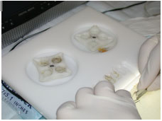
Graft drying is known to be one of the most detrimental factors influencing graft survival, and this system helps reduce the risk of graft drying by maintaining hydration during both the dissection and placing phases of surgery. Many clinics dissect grafts on a dry surface, place them in Petrie dishes, and then transfer grafts to a gloved hand -subjecting them to unnecessary handling and the risk of drying. The use of the dissection kit decreases graft handling, and decreases the risk of graft dehydration.
Dr. Keene sought a way to improve ergonomics and magnification over the commonly used microscopes used by hair transplant clinics. While seeking an ergonomically improved method of graft magnification, Dr. Keene worked with an engineer to co-develop the video magnification system, which not only offers ergonomic advantages, but also allows the doctor the opportunity to see every graft as it is being dissected. This also provides her the opportunity for maximum oversight and quality assurance before the grafts are placed!
As they are dissected, the grafts are stored in chilled lactated ringers in a dissection kit that Dr. Keene designed to maintain optimal graft hydration throughout the surgery. The graft receptacles can be transferred directly to the doctor’s or assistant’s finger for placing, without additional graft handling.